Keratoconus Treatment & Cornea Eye Surgery
Cornea care and keratoconus treatment
At Nandadeep Eye Hospital, our dedicated cornea eye clinic provides comprehensive care for a wide range of corneal conditions. We focus on accurate cornea diagnosis and offer advanced cornea treatment options for irregular disorders and progressive keratoconus. Led by experienced cornea specialists, our team creates personalized treatment plans ranging from early keratoconus diagnosis to advanced procedures such as keratoconus surgery and cornea transplant, when clinically required.

The unique features of the Cornea Department at Nandadeep Eye Hospital
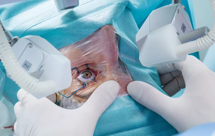
1. Advanced Operation Theater and Experienced Surgeons
- The Cornea Department boasts a state-of-the-art operation theater equipped with cutting-edge technology. Highly experienced ophthalmic surgeons perform intricate corneal procedures, ensuring precision and safety for patients.
- These skilled surgeons specialize in various corneal surgeries, including corneal transplants, lamellar keratoplasty, and other vision-restoring procedures.
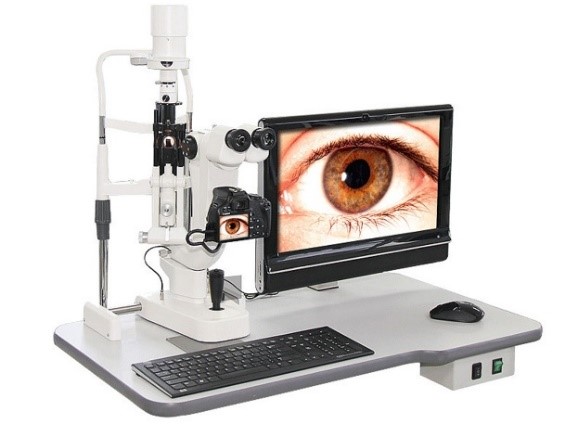
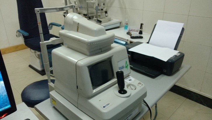
2. Diagnostic Equipment
- Nandadeep Eye Hospital invests in advanced diagnostic equipment specifically tailored for corneal evaluations. These tools allow accurate assessment of corneal health, shape, and thickness.
- High-resolution corneal topography, specular microscopy, and anterior segment optical coherence tomography (OCT) aid in diagnosing conditions like keratoconus, corneal dystrophies, and infections.
3. Unique in Southern Maharashtra:
- Nandadeep Eye Hospital stands as a pioneering institution in Southern Maharashtra, offering specialized corneal care. Patients from across the region seek treatment here due to its reputation for excellence.
- The hospital’s commitment to providing comprehensive corneal services sets it apart, making it a beacon of eye care in the area.
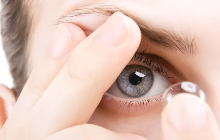
4.Specialty Contact Lens (CL) Department
The hospital’s CL Department caters to patients with various corneal conditions. Here are the specialty lenses offered:
- Scleral Lenses: These large-diameter lenses vault over the cornea, providing comfort and visual improvement for irregular corneas (e.g., keratoconus).
- RGP (Rigid Gas Permeable) Lenses: Ideal for correcting refractive errors and managing corneal irregularities.
- Rose K Lenses: Specifically designed for keratoconus patients, offering optimal fit and vision.
- Soft Toric Lenses: Used for astigmatism correction, especially when combined with other corneal conditions.
- Bandage Contact Lenses: These therapeutic lenses protect and promote healing in corneal abrasions or erosions.
The CL Department ensures personalized fittings, regular follow-ups, and patient education for successful lens wear.
Our hospital caters to a wide range of corneal diseases
Corneal Opacity
Corneal Opacity
- Blurred or cloudy vision.
- Sensitivity to light (photophobia).
- White or gray spots on the cornea
Keratoconus
Keratoconus diagnosis
- Blurred or distorted vision.
- Increased sensitivity to bright light and glare (especially at night).
- Frequent changes in eyeglass prescriptions.
Corneal Ulcers
Corneal Ulcers
- Red, watery, and bloodshot eye.
- Severe eye pain.
- Pus or other eye discharge.
Post-Traumatic Treatment
Post-Traumatic Treatment
- Symptoms related to trauma or injury, which can vary widely depending on the specific incident.
Acid or Alkali Burn
Acid or Alkali Burn
- Intense eye pain.
- Redness and swelling
- Blurred vision.
Stevens-Johnson Syndrome
Stevens-Johnson Syndrome
- Painful red or purplish rash, often starting on the face and spreading.
- Blisters or sores on the mucous membranes (including the eyes, mouth, and genitals).
- Eye redness, tearing, and sensitivity to light.
Foreign Body (Superficial and Deep)
Foreign Body
- Irritation.
- Sensation of something in the eye.
- Tearing.
Allergic Conjunctivitis
Allergic Conjunctivitis
- Itchy, red, and swollen eyes.
- Watery discharge.
- Sensitivity to light.
Severe Dry Eye
Severe Dry Eye
- Persistent dryness and discomfort.
- Redness.
- Blurred vision.
Sjogren Syndrome
Sjogren Syndrome
- Dry eyes and mouth.
- Fatigue.
- Joint pain.
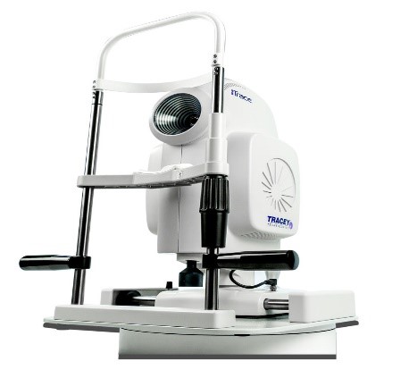
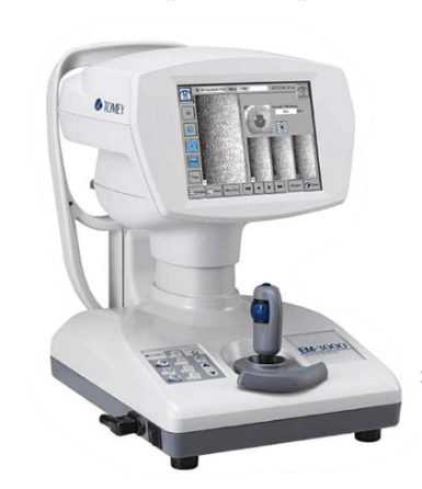
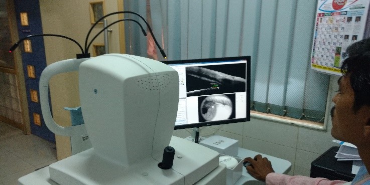
Investigations and diagnostic services available at our hospital
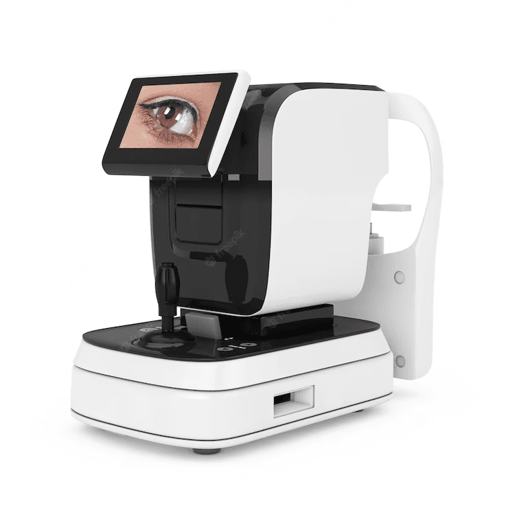
- Corneal Topography: A corneal topography test provides detailed maps of the cornea's shape and curvature and enables detection of corneal diseases.
- Pachymetry: Measurement of corneal thickness using ultrasound.
- Aberrometry: Measures the optical aberrations of the eye.
- Meibography: Used for dry eye work up
- Specular Microscopy: Study of the corneal endothelium (cells that maintain corneal clarity)
- Anterior – Segment OCT: Optovue helps to identify the depth of corneal scarring/lesions on the cornea, corneal thickness, anterior chamber details and any foreign body in cornea can be investigated.
- Anterior segment digital photography: Photographic documentation of various corneal conditions for baseline documentation, and monitoring effects of therapy as well as to monitor progression of the disease in any ectatic disorder.

Keratoconus treatment and cornea care at our eye hospital
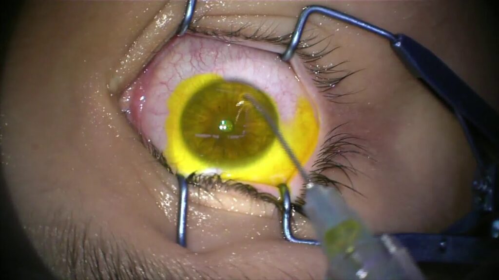
1. Surgical Treatments
Corneal Transplants (Keratoplasty):
Nandadeep Eye Hospital provides advanced corneal transplant procedures. These surgeries involve replacing a damaged or diseased cornea with a healthy donor cornea.
Types of corneal transplants include:
- Penetrating Keratoplasty (PKP): Full-thickness corneal transplant.
- Deep Anterior Lamellar Keratoplasty (DALK): Replaces only the anterior layers of the cornea.
- Endothelial Keratoplasty (DSAEK/DMEK): Replaces only the innermost endothelial layer.
C3R (Corneal Collagen Cross-linking):
- It’s a treatment for keratoconus.
- Strengthens the weakened corneal structure by inducing cross-links between collagen fibers using riboflavin (Vitamin B2) drops and UV light.
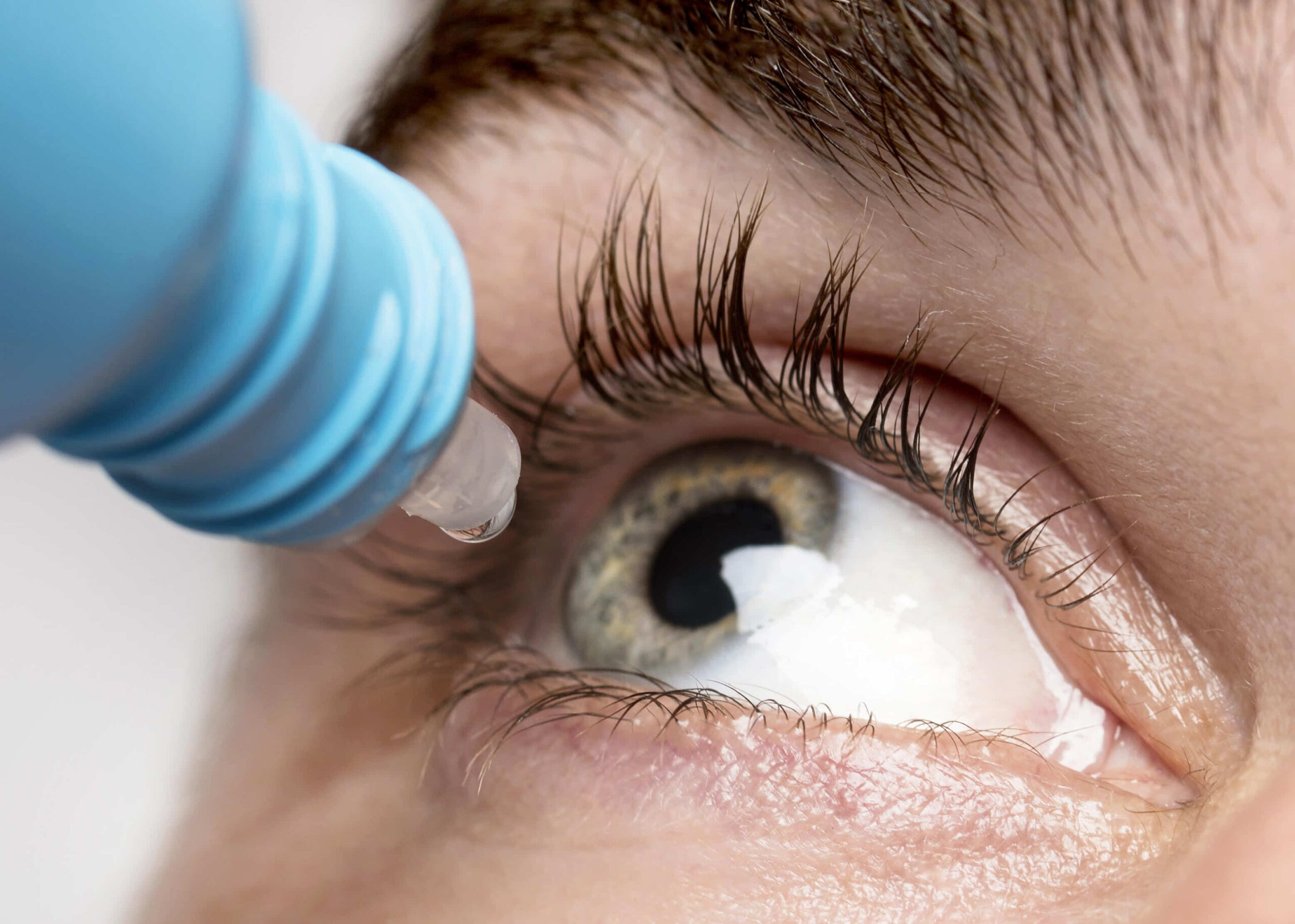
2. Medical Treatments:
Preservative-Free Artificial Tears:
- Used to alleviate symptoms such as dryness, discomfort, and itching.
Topical Immunosuppressive Treatment:
- May be prescribed for specific corneal conditions.
3. Non-Surgical Treatment:
Contact Lenses:
Nandadeep Eye Hospital offers specialized contact lenses for various corneal conditions:
- Scleral Lenses: Ideal for irregular corneas (e.g., keratoconus).
- Soft Contact Lenses: Used for conditions like dry eye or corneal irregularities.
- Rigid Gas Permeable (RGP) Lenses: Correct vision and improve comfort.
- Hybrid Lenses: Combine features of soft and RGP lenses.
- Custom-Made Lenses: Tailored to individual needs.
Frequently Asked Questions
The recovery occurs within hours of the death of the donor. The entire eye, called the globe, may be surgically removed (enucleated), or only the cornea may be excised in-situ and placed in storage media.
Practically anybody from the age of 1 year. There is no upper age limit. People who wear spectacles, who have undergone cataract surgery, diabetics, and those who have hypertension can donate their eyes. People blind from retinal or optic nerve disease can also donate their eyes. The ultimate decision about usage for transplantation will be made after evaluation.
Yes
Motivate the next of kin of the deceased person to donate their eyes. Eyes need to be collected within 6 hours of death so call your nearest eye bank at the earliest. You are authorized to donate the eyes of your beloved relatives at the time of their death, even if a pledge for donation has not been made earlier by the deceased.
- Keep the eyes of the deceased closed and cover it with moist cotton.
- Switch off the overhead fan.
- Raise the head end of the body by about 6 inches, if possible – to decrease the incidence of bleeding during the removal of the eyes.
- If possible, instill antibiotic eye drops periodically.
A call for donating eyes sets in motion the Eye Bank machinery and a team is dispatched within 15 minutes. At the site, the eyes of the deceased are enucleated without causing any disfigurement. This procedure takes about 10-15 minutes.
- Death from unknown cause.
- Death due to infectious caused viz. Rabies, syphilis, infectious hepatitis, septicemia, AIDS, corona.
- Any frank ocular infection, previous refractive surgery.
- Cancer in any part of the body.
No. Only the cornea and sclera can be transplanted. However, the entire eyeball is enucleated, to enable the corneo-scleral disc to be fashioned surgically in a sterile environment.
No. The gift of sight is made anonymously.
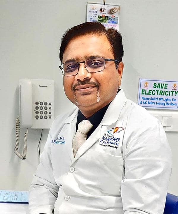
Our Cornea and Keratoconus Treatment Specialist

Dr. Sourabh D. Patwardhan
Phaco-Refractive-Vitreoretina- Glaucoma specialist FRCS (UK), MS (AIIMS), DNB, MNAMS, FICO


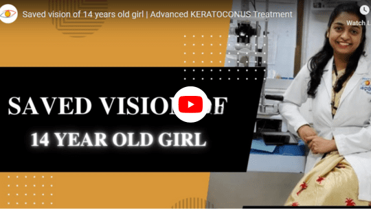
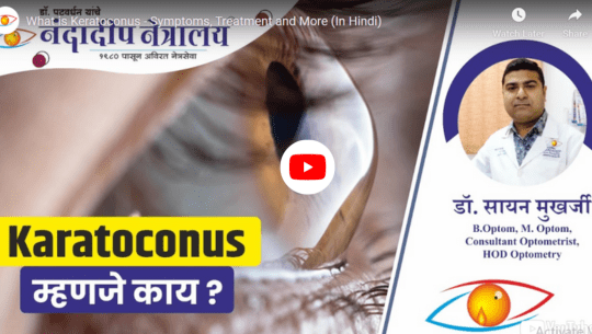
Why Choose Nandadeep Hospital ?Find Out the Latest Surgery Options and Types of Lenses Used for Cornea & Keratoconus Treatment
Explore our informative videos showcasing the latest advancements in Cornea & Keratoconus treatment. From minimally invasive techniques to cutting-edge procedures, we provide in-depth insights into the innovative surgical approaches available for effectively managing Pediatric and preserving your vision.